
Ultrasound may determine which premature babies can avoid damaging ventilation support
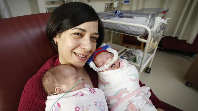
MELBOURNE doctors have for the first time captured moving pictures of what happens in the lungs of newborns as they take their first breath.
VIC News
Don't miss out on the headlines from VIC News. Followed categories will be added to My News.
LITTLE is known about the momentous instant in life when babies take a first breath.
Now, for the first time, Royal Women’s Hospital doctors have captured moving images of the lungs of newborns as they empty of amniotic fluid and fill with air for that initial gasp of oxygen.
The researchers hope that this safe and quick ultrasound technique, used in the first critical minutes after birth, can diagnose lung problems in premature babies.
DISCOVERY MAY PREVENT BABIES DEVELOPING CHRONIC LUNG DISEASE
BABY DIES AFTER REPORTED TWO-HOUR WAIT IN SUNSHINE HOSPITAL
This could help to better tailor treatment and prevent unnecessary use of ventilation, which can be damaging.
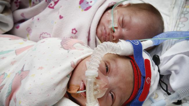
The team, in co-operation with The Ritchie Centre at the Hudson Institute of Medical Research, took ultrasound images of 115 full-term newborns. Images were taken of 28 babies before their first breath.
A series of six ultrasounds — taken from the first minute after birth through the first 24 hours — found there were significant changes in the lungs after just seven breaths, but that it took an average of four hours for all liquid to disappear. The study was published in the journal, Resuscitation.
Neonatologist Dr Doug Blank, the lead researcher, said categorising what happened in the lungs of healthy, full-term infants during the transition to the outside world provided a benchmark for detecting and treating respiratory problems in the immature lungs of premature babies.
Dr Blank said at least half of babies born at less than 29 weeks’ gestation would need mechanical breathing support and the delivery of surfactant to the lungs to help prevent the air sacs collapsing. This can be lifesaving, but if infants can remain stable on the gentler CPAP oxygen support, they are more likely to survive and avoid long-term lung damage.
“You’ve got this great treatment for the babies who need it, but it can be damaging for the babies who don’t need it,” Dr Blank said.
“I’m hoping this lung ultrasound can help us distinguish between these two groups of babies in the delivery room.”
About 50 preterm babies of less than 30 weeks’ gestation, born at The Women’s or Monash Women’s, will be recruited to an observational study of the use of ultrasound as a diagnostic tool to improve outcomes for the one in eight babies born prematurely.
“The lungs are connected to how the brain develops,” Dr Blank said.
“The more pristine the lungs are at the time we send an extremely premature baby home, the more likely they’ll have a good developmental outcome for things like learning and walking.”
Join the conversation

‘Angels’: Hero Coles workers save mum’s life
A mother of three has opened up on how her life was saved after she suddenly collapsed while doing a routine supermarket shop.
Read more
Wildlife and traffic fears over music festival
Wildlife advocates fear a weekend music festival in central Victoria will endanger native animals including rescued joeys.
Read more