We found the ‘silver bullet: Instant cancer detection on a glass slide
A smart microscope slide that could hasten cancer detection has ‘unlimited potential’.
A smart microscope slide developed by researchers at Melbourne’s La Trobe University could make early- stage cancer detection significantly quicker and easier, which could be key to increased rates of survival.
The scientists harnessed nanotechnology to develop the NanoMslide, the world’s first smart microscope slide. La Trobe physicist and professor of optics Brian Abbey and his university colleagues discovered that incorporating a silver membrane with nanometre apertures into a conventional slide can induce marked colour differences between cancerous and healthy tissues when viewed through a standard microscope.
“Basically, whatever sample is put on the surface creates these bright, vivid colours,” Abbey says. “The slide changes colour depending on the chemistry of the sample.”
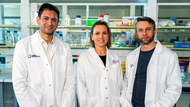
He has teamed up with Belinda Parker, a breast cancer specialist at the Peter Macallum Cancer Centre, to develop and validate the technology for commercial use, and they hope a commercial product will be available within five years. “We are manufacturing the slides here in Melbourne at the moment,” he says. “In collaboration with the Australian National Fabrication Facility, we’re in the process of scaling up our manufacturing for clinical- scale validation of the technology.”
Cancerous tissue has traditionally been identified by staining a mounted sample with dye to highlight abnormalities. That method, which has been around for more than a century, is slow and it can be inaccurate, particularly in early-stage cancers where only a few abnormal cells are present and visible differences in tissue structure can be minimal.
The NanoMslide’s ability to highlight even minor abnormalities with clear colour differences promises a significantly more accurate diagnostic process, Abbey says.
“In many cases, only a small number of abnormal cells are present, and there is a lack of effective and reproducible markers that work reliably across various very early-stage cancers,” he says. “This gap leads to higher rates of both false positives and false negatives during these critical early stages of detection.
“We are currently validating the ability of these slides to provide reliable detection of cancer versus non-cancer by testing them with a larger and more diverse set of patient tissue samples. The early results look promising.”
The NanoMslide does not require extra specialist pathology personnel or equipment, Abbey says, and it speeds up the process of cancer detection. Using the slide, pathologists can diagnose tissues directly at the microscope, without needing extra staining. In the future, this technology could give real-time feedback on whether tissues are cancerous, even while patients are in the operating theatre.
Abbey says he and his team are prioritising early-stage cancers and they are already looking beyond breast cancer to investigate the NanoMslide’s use in the detection of skin, colorectal and lung cancer as well. The tool’s potential is unlimited, he says.
“We describe this as the world’s first smart microscope slide, and it’s already being used for a huge range of different applications,” he says. “While we’ve concentrated mainly on cancer detection, these slides can be used to analyse almost any type of sample. They reveal the chemistry through unique colour changes, offering a capability that no other technology currently provides. This means there are virtually no limits to what these smart microscope slides could be used for.”

Adelaide’s vision for a university of the future
Adelaide University is already set to be Australia’s largest domestic educator: it will have more than 70,000 students and a projected domestic student base in excess of 70 per cent.

Universities must be top of agenda for growth
Cutting immigration robs the nation and the tertiary education sector of a source of revenue.