Oxford scientists: How we made the AstraZeneca vaccine
Catherine Green and Sarah Gilbert, the Oxford scientists behind the AstraZeneca vaccine, detail their painstaking work to crack Covid.

We were never searching for, hunting for, or finding a vaccine. Vaccines are not found. Even the world’s first vaccine, against smallpox, was the result of a carefully thought-out line of reasoning, not a happy accident.
Vaccines are first designed, based on scientific knowledge accumulated over decades and detailed study of the pathogen. Then they are made, carefully and painstakingly, by teams of people following strict rules and then tested for safety and efficacy.
The techniques we used to make the Covid-19 vaccine range from tried-and-tested methods that a 16-year-old science student might recognise, to newly developed, state-of-the-art technology.
The work was fiddly and exacting. The people, often manipulating tiny quantities of precious substances and working under layers of protective gear, were meticulous, systematic and dedicated. We were not completely unprepared: we had been thinking about, and trying to prepare for, Disease X for some time. But one thing we had not thought about was this: how do you fight a pandemic, when you are in a pandemic?
When it comes to making any adenovirus-vectored vaccine, which is what our Covid-19 vaccine is, I always tell people it is a bit like making bread. Partly because making vaccine from starting material – which is unique to adenovirus-vectored vaccines – is like making sourdough from a starter.
Here, step by step, is how we made (not found) the vaccine now being rolled out across the world.


STEP 1:
Make the starting material
We normally allow three to four months to make a starting material in the Clinical Biomanufacturing Facility using our classic method, but this time we were going to be attempting a new, more rapid method. At this point, my most optimistic estimate was that we might have a starting material ready by mid-March 2020, and the vaccine ready for clinical trials by the end of July. To hit these dates everything would need to go perfectly.
The rapid method, which we had never used before, would need to work. The team would need to do double shifts and come in on weekends. And we would need to proceed through the process “at risk” – in other words moving forwards to the next stage of the process before all testing was complete on the previous stage.
It’s important to be clear here that the risk is not to the end product, the vaccine itself, but to our time, effort and finances: if a problem is identified during testing, some of our work has been futile and we have to back up and repeat a step. In the end our first batch of starting material was ready, as we had hoped, by March 17, and the vaccine was ready for trials by April 22, much earlier than we had originally hoped.
On Tuesday, January 28, 2020, the pre-GMP team kicked off the rapid method to make the starting material by combining synthetic DNA that coded for the spike protein with a basic, all-purpose vaccine prepared earlier.
Now they hopefully had a sequence of DNA that coded for the Covid-19 vaccine. But virus DNA outside of a living cell is just an inert chemical. To allow the virus to assemble and replicate itself, the DNA needed to be inserted into living cells, which would become little microfactories, churning out copies of the adenovirus vaccine.
The method we use for inserting naked DNA into a human cell is called transfection and has been used for decades. The external membrane of human cells is made from a lipid (fatty) layer: you can imagine it like the outside of a washing-up-liquid bubble. This acts as a boundary between the substances inside the cell (proteins, chemicals, the cell’s own DNA) and the nutritious broth, or growth medium, that the cells are grown in.
The DNA for the vaccine cannot get across that membrane into the cells. So we put it in a solution that coats it in a bubble, similar to the lipid layer.
When the DNA in its solution is mixed with the cells, the two lipid surfaces can fuse, like when two small washing-up-liquid bubbles fuse to become one big bubble. The DNA is now inside the cell, where it provides the instructions for the cell to make lots of copies of the adenovirus.
But by February 17, nearly three weeks later, we could not detect virus particles in any of the dozens of preparations we had made. The rapid method had failed. So, on February 20, we activated plan B: the classic method, but faster.
We transfected the cells. After just one day of incubation, we diluted them in order to separate them into individual cells, and introduced each of these individual cells into a new, uninfected culture. Each single infected cell would then produce an independent preparation of the vaccine, which could be expanded and tested to check it was completely correct and homogeneous – a true clone.
By early March we had picked a favourite clone that was genetically perfect and proliferating particularly well: its name was D8.
We needed enough starting material to seed the manufacturing process many times over, so we now set about making a much larger preparation of D8.
In total we ended up making about 300ml of this precious fluid – enough to seed the manufacture of every dose of the Oxford vaccine, at least three billion doses, all born from this same starting material.
STEP 2:
Use the starting material to produce the required quantity of vaccine
By February the new coronavirus had seeded itself in dozens of countries and we had learned that people with the virus could be asymptomatic and be silently transmitting for days – the world was going to need this vaccine as soon as humanly possible.
We decided to proceed “at risk”, moving to the next stage of the process before all testing was complete on the previous stage. If the starting material failed any of its tests, we would have to throw out anything we had made from it.
To make the vaccine, you need to introduce the starting material into the human cells that will act as the virus-production factories.
On March 6, two precious vials of specially adapted cells were introduced into a small conical flask of growth medium. Over the next two weeks, the cells grew exponentially: that is, in the first day or so they doubled, then in the next few days they doubled again, and so on. Two weeks later we had almost 10 litres of cell culture, ready to receive the virus starting material.
At the same time as our production cells were growing exponentially in the lab, so were case numbers out in the world. And the consequences for our daily lives were doubling and redoubling too. I needed the members of my small team not to get the virus, and definitely not to pass it on to their colleagues – nobody else could do this work. It was impossible to get hold of hand sanitiser so we made our own-brand batch, using lab chemicals according to the WHO recipe. It was thin and smelt of alcohol but it would work. On March 23 we inoculated our 10 litres of culture using about 10 millilitres of our starting material. This was a big moment. We were putting the bread in the oven, and then we would just have to wait to see whether it was going to rise.
Two days later, our bread was ready. Our production cells had produced lots of adenovirus vaccine particles but not yet burst. The harvesting could begin.
STEP 3:
Purify the vaccine
In this step, we get rid of all of the bits of the human cells in which we have grown our viral vaccine, and any badly formed non-functional viral particles, leaving us with pure vaccine. We want the bread, not the tin we baked it in.
We had another difficult choice to make about the method we would use and decided to split the batch. Two of the seven flasks would be purified with a new method, and five with the traditional one. We started with the traditional method.
At the time of the harvest, most of the adenovirus vaccine particles are still inside the production cells. The cells are relatively large so they can easily be separated from the nutrient broth by a low-speed centrifugation. The culture of vaccine-containing cells is simply transferred to sterile bottles, which are spun at 3000rpm in a centrifuge – effectively just a large, high-powered salad spinner.
All the cells containing the vaccine form a pellet at the bottom of each bottle, and the broth can simply be poured away.
We need the vaccine to be pure. We don’t want traces of the production cells in it. To get the vaccine out from the pelleted cells, we add a lysis buffer (a solution of salts and detergents) and “pop” the cells by freezing and thawing them three times. As the cells freeze, the liquid inside them forms into ice and expands, bursting the cells’ external membrane. The contents – including the virus particles – leak out into the buffer solution. We then thaw and recentrifuge and because the vaccine particles are very small they stay in the solution while the broken bits of cell form a new pellet at the bottom of the tube. Now we have a solution called clarified cell lysate – this contains all the properly formed virus particles, but also any empty adenovirus shells that have not formed properly and will be useless as vaccines, as well as other soluble material that came out from the cells when they burst.
To purify the active vaccine, we use the fact that the particles we want have a different density from all the bits we don’t want. We make two salty solutions of different densities and carefully layer them inside a specially strengthened test tube. Then we carefully layer the clarified cell lysate containing our vaccine particles on top, then centrifuge the cocktail at a very high speed.
After two hours of spinning, all the properly formed vaccine particles are gathered together in a single band within the tube, with the empty, unwanted particles and other bits we don’t want in another layer higher up. We collect the liquid containing our vaccine particles using a needle and syringe inserted through the side of the tube, then repeat the centrifugation process and carry out a final purification step.
Purification is always a nervous few days in the clean room. The vaccine is all contained in a few flasks, then a few tubes, and finally a single bottle or bag, and a mistake at any point can jeopardise everything. You need a steady hand and a calm temperament. Fortunately the team had both.
On March 27 a team member opened the centrifuge, removed the tubes that had gone through the traditional purification method and took a photo that showed an enormous fuzzy cloud of vaccine right in the centre of the tube. Later that day she emailed it to the whole team, with the subject line “Look at these babies!” Someone emailed back, only half-joking: “This might just be the fuzzy band that saved the world.”.
The whole team was on a high. I think this was the moment we believed we might pull this off.
Our first batch of vaccine – less than 500ml – was put into a sterile bag and into the freezer, ready for the next step: putting the vaccine into vials.
Then we went back to process the second batch using the new method. We had apparently good yields and the first step of the purification went as expected.
But on the evening of April 8 we had the devastating news the material that was coming off the final step was behaving strangely. Instead of the expected perfectly clear solution, the liquid emerging was cloudy.
We knew this was not a good sign. We had never seen this before and no one really knew what to do. It was late at night so we agreed that we should put everything safely in the fridge, everyone should go home and try to get some sleep, and we would approach it with fresh brains in the morning.
The next morning we ran some tests. Very little vaccine could be detected and by that evening it was clear that it was hopeless. Our best guess was that the virus particles had all clumped together somewhere unexpected at the final stage and been accidentally discarded.
I don’t think I can explain how awful this felt for the team. They knew how important this was and they felt personally responsible for the failure. That night I gave everyone a pep talk: they had already done one impossible thing, I told them, creating the first batch in record time. Asking for two impossible things was just too much.
STEP 4:
Fill the vials
Because we are going to put it into people’s arms, the final vaccine product of course must be completely sterile. Microorganisms are all around us, in the air, in the soil and on our bodies. So you can fill vaccine into vials only in the most stringently sterile environment.
Because we only make small batches, the simplest and most cost-effective way for us to fill vials is manually, inside an isolator which is itself inside a clean room.
The isolator is a sealed box, the width of the room, into which completely purified air is pumped to keep it sterile. One side is a glass screen with two pairs of sealed gloves set into it so that the operators, sitting side-by-side outside the box in their full protective gear, can handle the materials and equipment inside the box without touching it with their hands.
There is a hatch at each end so that wrapped, sterilised materials and equipment can be placed inside the box from one end, and filled sterile vials can be removed from the other. Vial filling is one of the most skilled jobs that we do. It is high-pressure work that is also boring, repetitive, delicate and tiring on the arms. The production team know how to handle it though and are very steady.
On March 28, we had deposited our first successfully purified batch in the freezer.
A couple of days later I did a radio interview in which our work was introduced as “the only story in the world”. My mind flashed to the lab. There was so much to do, and so much that could still go wrong. Several members of the team were having to self-isolate as they lived in shared houses with a housemate who had symptoms. Being the only story in the world felt like quite a heavy weight to bear.
Thursday, April 2, was fill day – always a big day. The whole team is involved as there is lots of checking, testing, monitoring, distribution and documentation to do.
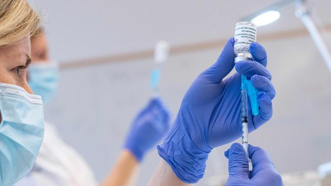
We removed our small bag of vaccine from the freezer, transferred it into the isolator and then pumped it through a series of filters with tiny pores that remove any bacteria. The final sterile filtered material was then ready for filling. Using a tiny, very precise syringe, a member of the team carefully placed approximately 0.5ml (a tenth of a teaspoon) of vaccine into each glass vial.
By the end of the day the team had filled 500.
STEP 5:
Label the vials, document the process, and test and certify the quality of the product
This is steady, behind-the-scenes work, checking everyone else’s work and ensuring that strict standards are met.
Labelling might sound like a simple job, but there are very comprehensive regulations involving a lot of documentation to ensure that products for clinical trials are properly labelled. A mistake could result in a wrong drug or a wrong dose, which could be very serious.
Documentation is necessary in order to get approval to go ahead with any trial. There are detailed protocols for everything, and detailed records kept of everything that happens.
Testing is not glamorous or exciting but it is obviously a crucial part of the process.
The final step is certifying that the batch produced is suitable for use in a specific trial. We got our final test results back on April 21 and the next day the batch was certified as ready for use. We were all exhausted.
We had a slice of socially distanced cake and went home to try to get some sleep.
Vaxxers: The Inside Story of the Oxford AstraZeneca Vaccine and the Race Against the Virus by Sarah Gilbert and Catherine Green; Hodder & Stoughton, $34.99
To join the conversation, please log in. Don't have an account? Register
Join the conversation, you are commenting as Logout