
A rare glimpse into Australia’s body part museum
A FASCINATING rare glimpse into the Canberra museum of body parts usually only open to medical students and living donors. WARNING: Graphic images.

ROGER* was a normal 26-year-old enjoying a night out with his mates when he died suddenly from cardiomyopathy, an inherited heart condition that he was unaware of. Amid the terrible grief of losing her son, Roger’s mother Fran* did something extraordinary and agreed to donate his enlarged heart to the Peter Herdson Museum so medical students could learn from it.
Fran made a phone call to Jane Dahlstrom, Professor of Anatomical Pathology at the Canberra Hospital, and explained her son was “was a really generous person, and I’m sure he would really like to do this.”
As Professor Dahlstrom relates this story back to me, she is clearly moved.
“I feel very blessed that people would even think about other people when they’re obviously suffering themselves,” she says.

The Peter Herdson Museum, which is housed at the Canberra Hospital, contains more than 1,200 specimens of human tissue. Some of them date back to the 1940s and 50s. Other organs in the Museum are contemporary specimens that have living donors who give their diseased organs to the collection after having them removed during surgery.

The third category is those samples like Roger’s heart that are donated by deceased people or their families for the benefit of Australian National University (ANU) medical students studying in both Canberra and the rural areas of NSW.
Professor Dahlstrom says a conversation like the one she had with Roger’s bereaved family can be heartbreaking and occasionally humorous too.
“We had a chat about how they would like the heart to be kept. Did they want the heart in a pot, or did they want the heart to be able to be touched by the medical students?” Professor Dahlstrom explains.
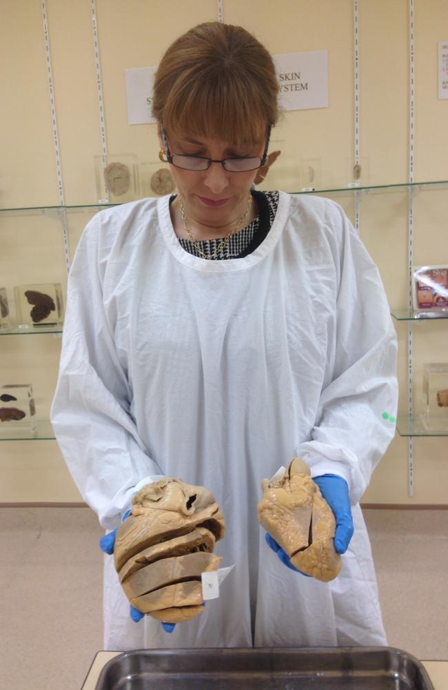
Fran replied that her son “wouldn’t mind the students to touch and feel him.”
“This is why this specimen is not potted,” Professor Dahlstrom says, adding that Roger “must have been a really lovely person.”
The Museum contains so-called “potted” specimens that are preserved in glass jars for the medical students to view and “wet” specimens that are preserved in 10 per cent alcohol for students to handle and observe as part of their education.
The facility is not normally open to the public but Professor Dahlstrom does frequently arrange viewings for living patients with organs displayed in the Museum or “perhaps the next-of-kin if the patient isn’t alive.”
She says this can be important because “they want to see it for themselves, and I think that can be helpful for closure.”

Sandy Cochrane is a fourth-year student at the ANU Medical School and says the Museum “allows us to see things in three dimensions that are very difficult to see in a living person and that can’t really be represented by a picture or by a drawing.”
At the same time Mr Cochrane says that seeing “large portions of people who had died and whose body parts had been preserved for our study” was confronting in first year pathology laboratory classes because “it takes a while to accept mortality.”
“I recall seeing a young woman [and] … finding that very challenging to accept that someone younger than me could have passed away,” he says.
Now Mr Cochrane is in his final two years of study, he’s far more accustomed to the human body. He shows me a cirrhotic liver specimen in the Museum. Because of examples like this one, Mr Cochrane says, he was well prepared when examining under the right rib cage of a living patient with this “common and serious and often preventable disease.”
“A cirrhotic liver is usually smaller than a normal healthy liver, and it’s full of little hard nodules, a bit like little hard frozen peas,” he explains.

These preserved museum specimens “helped give me a mental picture of what it was that I was feeling in that person,” Mr Cochrane continues.
Dr Elizabeth Paver, a first-year anatomical pathology registrar at the Canberra Hospital, says the Museum gives trainee medical students and trainee doctors an important opportunity to build up their knowledge of “the really rare and the really unusual conditions as well the more common diseases.”
In answer to which specimen struck her most, Dr Paver shows me a liver that has sustained a gunshot wound.

“Seeing the damage that it’s done, but also the liver’s capacity to regenerate around where the shrapnel went through … it just illustrates how amazing the body is in its capacity to deal with these sorts of traumas,” she says.
“All of these specimens, they all have a story behind them, and the people that have kindly donated them have a story,” Dr Paver says.


*Names have been changed.
Ginger Gorman is an award winning print and radio journalist, and a 2006 World Press Institute Fellow. Follow her on twitter @freshchilli

Terror as man dies from the black plague
While you may assume the horror of the ‘black death’ is confined to history books, a man’s sudden death from the plague has changed everything.

World-first move for gay blood donors
Rules that have restricted members of the LGBTQIA+ community from donating blood in Australia for decades have just been lifted.

Psychedelic drugs trial gets green light
A major Aussie university and ASX-listed company will deploy psychedelic drugs to treat binge-eating in a world-first clinical trial.